
Periodontal Gingival Graft Treatment: A Case Of Canine Recession
Explore gingival graft treatment for canine recession completed at SOL Dental Arts in Maspeth, Queens.
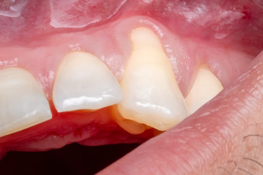
Gingival Graft For Canine Recession
Soft tissue treatment addressing gum recession around a canine tooth to improve protection and esthetics.
This gingival graft case at SOL Dental Arts in Maspeth, Queens addresses recession around a canine tooth with soft tissue treatment designed to improve root protection, reduce sensitivity concerns, and enhance gumline appearance. When gum recession exposes the root surface, the issue can be both functional and cosmetic. A grafting procedure may be recommended to strengthen the area, improve tissue coverage, and create a more stable, natural-looking gum contour.
This type of case is especially important when the patient wants to address recession early and protect long-term periodontal health while also improving smile esthetics.
What Was Done?
- Evaluation of canine recession
- Periodontal soft tissue grafting procedure
- Root coverage and gumline contour improvement
- Follow-up healing assessment
How We Do It
Canine recession can increase sensitivity, expose root structure, and create asymmetry in the smile. A graft helps improve tissue protection and esthetics.
Frequently Asked Questions
Why Is Gum Recession On A Canine Important To Treat?
Canine teeth are prominent in the smile, and exposed roots may affect both comfort and appearance.
Is A Gingival Graft Only Cosmetic?
No. It can also help protect the root and improve long-term tissue health.
Can Grafting Improve Smile Symmetry?
Yes. Better gum coverage can improve the balance of the gumline.

Related Treatment Resources
For treatment context, explore gum recession treatment, dental cleaning and exams, gum recession and gingival grafting, and tooth-colored bonding for a front tooth cavity.
Ask About A Similar Result
Every smile and treatment plan is individual. Meet our dentists or request an appointment to discuss what may be appropriate for you. You can also call our office. Individual results vary.
Diagnostic Evaluation & Anatomical Vulnerability Of Canines
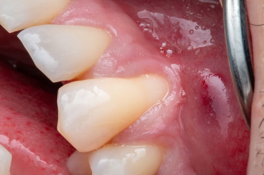
Canines occupy a prominent, high-stress position in the dental arch, guiding jaw movements and absorbing significant lateral forces during chewing. Due to their prominent position in the jawbone and the thin layer of overlying bone and tissue, upper and lower canines are naturally prone to gingival recession. Aggressive toothbrushing habits, anatomical prominence, and clenching forces can easily wear away fragile marginal tissue, exposing sensitive dentin and vulnerable root cementum.
At SOL Dental Arts in Maspeth, NY, our diagnostic evaluation begins with a detailed periodontal risk assessment. Our doctors utilize low-dose digital x-rays to measure interdental bone levels and evaluate underlying root stability. By assessing tissue thickness and probing depths around the recession site, our team identifies the exact cause of tissue breakdown before recommending surgical intervention, ensuring that the root cause is addressed alongside soft tissue coverage.

Key Factors In Evaluating Canine Soft Tissue Deficits
- Assessing the width of attached keratinized tissue to determine long-term periodontal resistance against mechanical wear
- Reviewing low-dose digital x-rays to verify interdental bone height and rule out underlying periodontal bone loss
- Checking static and dynamic chewing guidance to ensure heavy lateral forces are not flexing the tooth
- Examining root surface integrity to rule out subgingival notch defects or active root decay before grafting
Advanced Grafting Techniques & Long-Term Periodontal Health
Restoring soft tissue coverage over an exposed canine root requires specialized periodontal techniques that build thick, durable keratinized tissue. Depending on anatomical needs, subepithelial connective tissue grafts or advanced acellular dermal matrices are positioned precisely over the cleansed root face. This donor tissue acts as a scaffold, encouraging surrounding blood vessels and collagen fibers to regenerate a healthy, attached gum margin.
During care at our practice in Maspeth, NY, our clinical team prioritizes patient comfort and delicate tissue management. The grafted site is anchored with micro-sutures to ensure graft stability during initial vascularization. As healing progresses, the newly reinforced gum tissue shields the exposed root from hot and cold thermal sensitivity, prevents root caries, and establishes a balanced, symmetrical gumline that frames the smile naturally.
Essential Care Practices Following Gingival Grafting Procedures
- Avoid brushing or flossing directly around the surgical site during initial healing to protect delicate tissue attachments
- Rinse gently with prescribed antimicrobial mouthwash to keep graft borders clean without mechanical irritation
- Consume a soft-food diet and refrain from pulling on the lip to inspect the healing gumline
- Attend scheduled follow-up appointments so our team can evaluate tissue healing, monitor graft integration, and remove sutures safely